
ICE: How to Read the Heart
Dawn of Zero-Fluoroscopy: ICE Mastery
A practical video series designed for electrophysiologists dedicated to achieving true zero-fluoroscopy ablation through advanced ICE interpretation.
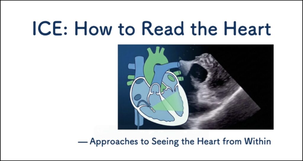
This series explores how intracardiac echocardiography can be used to better understand cardiac anatomy, catheter position, and procedural decision-making from inside the heart.
Available Now
Coming Soon
Modules currently in production

A knowledge hub for cardiac rhythm disorders and safer electrophysiology.
Refund Policy:
Due to the nature of digital video content, all sales are final and non-refundable.
Legal Notice (Specified Commercial Transaction Act)
Seller: NE LLC
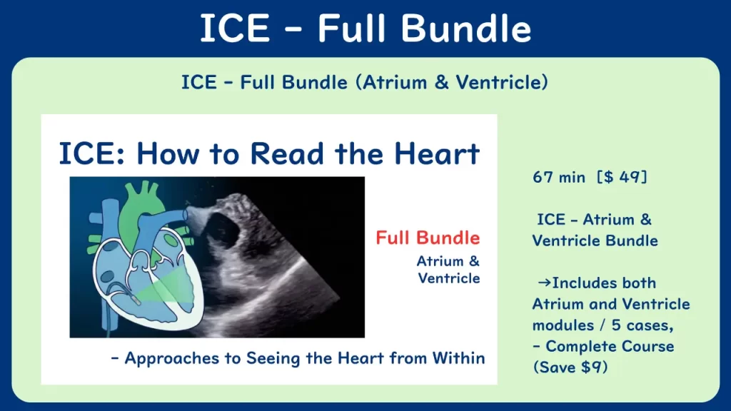
ICE: How to Read the Heart _ Full Bundle
For detailed descriptions of each video, please see the individual Atrium and Ventricle sections below.
After completing your purchase, you will be redirected to your private viewing page.
Please bookmark your private viewing page, as it will be your primary access point for future viewing.
Refund Policy:
Due to the nature of digital video content, all sales are final and non-refundable once purchase is completed.
A knowledge hub for cardiac rhythm disorders and safer electrophysiology.
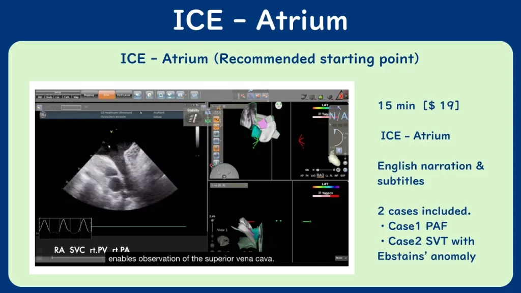
ICE: How to Read the Heart _ atrium
A practical, case-based video that shows how atrial structures truly appear from intracardiac perspectives — including Doppler-based flow assessment as applied to atrial imaging.
- Recognize atrial anatomy from real ICE views
- Understand atrial anatomy
- Apply Doppler-based flow assessment within atrial structures
- Build visual confidence for EP procedures
After completing your purchase, you will be redirected to your private viewing page.
Please bookmark your private viewing page, as it will be your primary access point for future viewing.
Refund Policy:
Due to the nature of digital video content, all sales are final and non-refundable once purchase is completed.
Who This Is For
Electrophysiologists, cardiologists, fellows, and imaging specialists
seeking visual clarity and spatial orientation using ICE during EP procedures.
You may find this valuable if:
- You perform ablation, device, or structural procedures
- You collaborate on imaging during EP cases
- You want to build pattern recognition, not only principles
- You aim to read ICE views with confidence
The Gap — Why This Training Exists
Knowing ICE is not the same as seeing with ICE.
Textbooks explain principles, but
visual pattern recognition develops only through repeated exposure to real views.
Challenges this video addresses:
- Textbooks rarely show procedural anatomy from within
- Spatial reasoning requires repeated visual patterns
- Most resources lack EP-specific imaging guidance
- Pattern recognition develops only through real-case observation
This series was created to bridge that gap.
What You Will Learn
By watching the series, you will learn to:
- Identify atrial structures from intracardiac perspectives
- Understand pulmonary vein and left atrial appendage views relevant to atrial imaging
- Apply Doppler-based flow assessment within atrial anatomy
- Build visual pattern recognition to improve spatial orientation during EP procedures
These are practical visual skills —
something textbooks alone cannot provide.
Curriculum Overview
This is the first video in the series, focused on atrial anatomy.
It includes Doppler-based flow assessment as applied to atrial imaging.
Topics covered:
- Atrial anatomy from intracardiac views
- Pulmonary vein & LAA imaging perspectives
- Doppler-based flow assessment within atrial structures
Why This Series Is Different
- Focused on procedural pattern recognition
- Views recorded during real catheter-based cases
- Clarity on how structures truly appear from within
- Narrated in English for an international audience
- Created by a clinician performing zero-fluoroscopy ablations
Not theoretical ICE.
Practical ICE — as used in electrophysiology.
About the Instructor
Toru Kawakami, MD — Electrophysiologist, Nagoya, Japan
Dr. Kawakami performs catheter ablation with a focus on radiation-free practice,
using ICE for procedural visualization, orientation, and safety.
This video series reflects his approach to seeing the heart from within
and teaching others to build the same visual confidence.
Purchase
ICE: How to Read the Heart — Video : Atrium
$19 USD — one-time purchase, on-demand streaming access
This video is designed as an educational resource for healthcare professionals.
It is not intended to replace clinical judgment or individualized medical care.
Taxes, VAT, or other applicable duties are not included in the listed price and
remain the responsibility of the purchaser.
You receive:
- Full access to Video : Atrium
- Case1 Normal heart
- Case2 Ebstein’s anomaly
- Doppler-based flow assessment included
- English narration & subtitles
Watch at your own pace —
pattern recognition grows through repetition.
After completing your purchase, you will be redirected to your private viewing page.
Please bookmark your private viewing page, as it will be your primary access point for future viewing.
Refund Policy:
Due to the nature of digital video content, all sales are final and non-refundable once purchase is completed.
FAQ
Q: Is Doppler included?
Yes. Doppler-based flow assessment is included as part of atrial imaging.
Q: Is this the complete series?
This is Video 1. Ventricular Anatomy will follow as Video 2.
Q: Is this suitable for fellows?
Yes, it helps connect anatomy, Doppler, and procedural imaging.
Q: Can I watch at my own pace?
Yes, access is streaming-based.
Q: How can I watch the video after purchase?
After completing your purchase, you will be redirected to your private viewing page.
Please bookmark this page, as it will be your primary access point for watching the video in the future.
You can watch the video anytime by accessing your bookmarked private viewing page.
Q: Are subtitles available?
Yes. English subtitles are available and can be turned on or off using the subtitle settings in the video player.
Additional language subtitles may be added in the future.
Q: What devices are supported?
The video can be viewed on desktop and laptop computers, tablets, and smartphones with a stable internet connection.
For the best viewing experience, we recommend using a desktop or laptop computer with an up-to-date web browser.
A knowledge hub for cardiac rhythm disorders and safer electrophysiology.
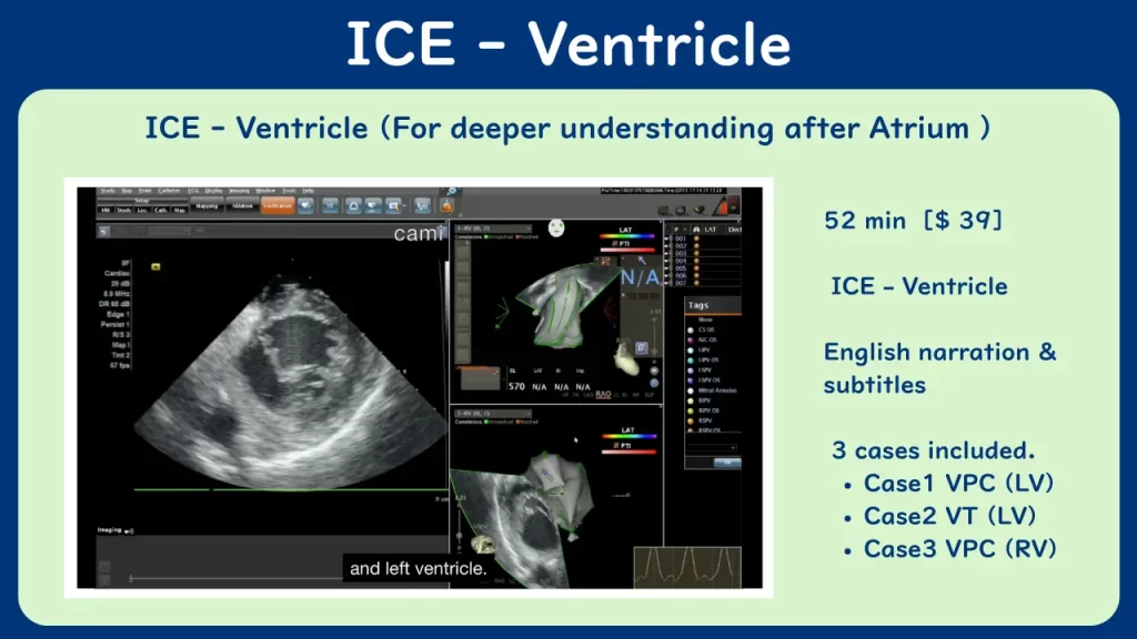
ICE: How to Read the Heart _ ventricle
A practical, case-based video that shows how ventricular structures truly appear from intracardiac perspectives.
- Recognize ventriclar anatomy from real ICE views
- Understand ventricular anatomy
- Build visual confidence for EP procedures
After completing your purchase, you will be redirected to your private viewing page.
Please bookmark your private viewing page, as it will be your primary access point for future viewing.
Refund Policy:
Due to the nature of digital video content, all sales are final and non-refundable once purchase is completed.
Who This Is For
Electrophysiologists, cardiologists, fellows, and imaging specialists
seeking visual clarity and spatial orientation using ICE during EP procedures.
You may find this valuable if:
- You perform ablation, device, or structural procedures
- You collaborate on imaging during EP cases
- You want to build pattern recognition, not only principles
- You aim to read ICE views with confidence
The Gap — Why This Training Exists
Knowing ICE is not the same as seeing with ICE.
Textbooks explain principles, but
visual pattern recognition develops only through repeated exposure to real views.
Challenges this video addresses:
- Textbooks rarely show procedural anatomy from within
- Spatial reasoning requires repeated visual patterns
- Most resources lack EP-specific imaging guidance
- Pattern recognition develops only through real-case observation
This series was created to bridge that gap.
What You Will Learn
By watching the series, you will learn to:
- Identify ventricular structures from intracardiac perspectives
- Build visual pattern recognition to improve spatial orientation during EP procedures
These are practical visual skills —
something textbooks alone cannot provide.
Curriculum Overview
This is the first video in the series, focused on ventricular anatomy.
Topics covered:
- Ventricular anatomy from intracardiac views
- Papirally muscles imaging perspectives
About the Instructor
Toru Kawakami, MD — Electrophysiologist, Nagoya, Japan
Dr. Kawakami performs catheter ablation with a focus on radiation-free practice,
using ICE for procedural visualization, orientation, and safety.
This video series reflects his approach to seeing the heart from within
and teaching others to build the same visual confidence.
Purchase
ICE: How to Read the Heart — Video : Ventricle
$39 USD — one-time purchase, on-demand streaming access
This video is designed as an educational resource for healthcare professionals.
It is not intended to replace clinical judgment or individualized medical care.
Taxes, VAT, or other applicable duties are not included in the listed price and
remain the responsibility of the purchaser.
You receive:
- Full access to Video : Ventricle
- Case1 VPC patient
- Case2 VT patient
- Case3 VPC patient
- English narration & subtitles
Watch at your own pace —
pattern recognition grows through repetition.
After completing your purchase, you will be redirected to your private viewing page.
Please bookmark your private viewing page, as it will be your primary access point for future viewing.
Refund Policy:
Due to the nature of digital video content, all sales are final and non-refundable once purchase is completed.
FAQ
Q: Is this suitable for fellows?
Yes, it helps connect anatomy, Doppler, and procedural imaging.
Q: Can I watch at my own pace?
Yes, access is streaming-based.
Q: How can I watch the video after purchase?
After completing your purchase, you will be redirected to your private viewing page.
Please bookmark this page, as it will be your primary access point for watching the video in the future.
You can watch the video anytime by accessing your bookmarked private viewing page.
Q: Are subtitles available?
Yes. English subtitles are available and can be turned on or off using the subtitle settings in the video player.
Additional language subtitles may be added in the future.
Q: What devices are supported?
The video can be viewed on desktop and laptop computers, tablets, and smartphones with a stable internet connection.
For the best viewing experience, we recommend using a desktop or laptop computer with an up-to-date web browser.
A knowledge hub for cardiac rhythm disorders and safer electrophysiology.
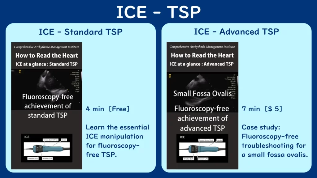
ICE: How to Read the Heart _ TSP
A concise, high-yield visual guide to ICE-guided transseptal puncture (TSP) without fluoroscopy.
Designed for fellows and junior EPs to master essential catheter manipulation and build clinical confidence in just minutes.
- ICE – Standard TSP → [Free] Learn the fundamental views and essential catheter manipulation for fluoroscopy-free TSP. ( 4 min )
- ICE – Advanced TSP → [$5] Case study: Fluoroscopy-free troubleshooting for a small fossa ovalis. ( 7 min )
After completing your purchase, you will be redirected to your private viewing page.
Please bookmark your private viewing page, as it will be your primary access point for future viewing.
Refund Policy:
Due to the nature of digital video content, all sales are final and non-refundable once purchase is completed.
About the Instructor
Toru Kawakami, MD — Electrophysiologist, Nagoya, Japan
Dr. Kawakami performs catheter ablation with a focus on radiation-free practice,
using ICE for procedural visualization, orientation, and safety.
This video series reflects his approach to seeing the heart from within
and teaching others to build the same visual confidence.
Purchase
ICE: How to Read the Heart — TSP
$5 USD — one-time purchase, on-demand streaming access
This video is designed as an educational resource for healthcare professionals.
It is not intended to replace clinical judgment or individualized medical care.
Taxes, VAT, or other applicable duties are not included in the listed price and
remain the responsibility of the purchaser.
You receive:
- Full access to Video : TSP
- Learn the fundamental views and essential catheter manipulation for fluoroscopy-free TSP
- Case study: Fluoroscopy-free troubleshooting for a small fossa ovalis
Watch at your own pace —
pattern recognition grows through repetition.
FAQ
Q: Can I watch at my own pace?
Yes, access is streaming-based.
Q: How can I watch the video after purchase?
After completing your purchase, you will be redirected to your private viewing page.
Please bookmark this page, as it will be your primary access point for watching the video in the future.
You can watch the video anytime by accessing your bookmarked private viewing page.
Q: What devices are supported?
The video can be viewed on desktop and laptop computers, tablets, and smartphones with a stable internet connection.
For the best viewing experience, we recommend using a smartphone with an up-to-date web browser.
A knowledge hub for cardiac rhythm disorders and safer electrophysiology.